Suche
Lesesoftware
Info / Kontakt
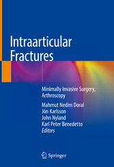
Intraarticular Fractures - Minimally Invasive Surgery, Arthroscopy
von: Mahmut Nedim Doral, Jón Karlsson, John Nyland, Karl Peter Benedetto
Springer-Verlag, 2019
ISBN: 9783319976020 , 419 Seiten
Format: PDF, Online Lesen
Kopierschutz: Wasserzeichen


Preis: 160,49 EUR
eBook anfordern 
Foreword
6
Preface
8
Preface
10
Preface
11
Acknowledgments
12
Contents
13
Part I: General Knowledge
17
1: Natural History of Bone Bruise
18
1.1 Introduction
18
1.2 Bone Bruise Classification
19
1.3 Bone Bruise Location and Mechanism
19
1.4 Clinical and Histological Findings
21
1.5 The Natural Course
22
1.6 Treatment
23
1.7 Conclusion
23
References
24
2: Arthroscopic Treatment Vs. Open Surgery in Intra-articular Fractures
26
2.1 Background
26
2.2 Articular Fracture Reduction
27
2.3 Associated Soft Tissue Injuries
27
2.4 Loose Bodies
28
2.5 Articular Degenerative Changes
28
2.6 Conclusion
29
References
29
3: Intra-articular Fractures: Principles of Fixation
30
3.1 Introduction
30
3.2 Classification
30
3.3 Unique Features of Intra-articular Fractures
33
3.4 Imaging of Intra-Articular Fractures
33
3.5 Basic Principles of Management of Intra-articular Fractures
33
3.6 Importance of Step-Offs/Gaps
35
3.7 Healing of Articular Cartilage
36
3.8 Conclusion
37
References
37
4: Intra-articular Fractures: Philosophy of Minimally Invasive Fixation
39
4.1 Minimally Invasive Fixation
39
4.2 Intra-articular Fractures
40
4.2.1 Description
40
4.2.2 Problems Related to the Treatment
41
4.2.3 Treatment Planning
41
4.3 Conclusion
42
References
42
5: Biologic Solutions for Articular Cartilage Healing
44
5.1 Introduction
44
5.2 Articular Cartilage Surgical Treatment
45
5.2.1 Reconstructive Procedures
45
5.2.2 Tissue Engineering and Scaffold-Based Procedures
46
5.3 Nonsurgical Articular Cartilage Treatment
49
5.3.1 Injections
49
5.3.2 New Injective Biological Approaches
50
5.4 Conclusion
50
References
50
6: Rehabilitation Principles Following Minimally Invasive Fracture Fixation
54
6.1 Introduction
54
6.2 Postsurgical Malalignment, Segment Length, or Joint Surface Inclination Changes
54
6.3 Healing Potential
55
6.4 Articular Surface Congruency
56
6.5 Potential Stress Shielding or Stress Riser from Fixation Hardware
57
6.6 Patient Expectations “Realistic or Not”
57
6.7 Optimizing Full Kinematic/Kinetic Chain Function
57
6.8 Patient/Client Understanding, the Importance of Therapeutic Lessons
58
6.9 Optimizing Metabolic Energy System Function
59
6.10 Repetitive Microtraumatic, Acute Isolated, or Polytraumatic Intra-articular Fractures
59
6.11 Pain
59
6.12 Gender, Genetics, Lifestyle, and Age
60
6.13 Therapeutic Exercise to Improve Function and Cognitive Appraisal: Psychobehaviors
61
6.14 Therapeutic Exercise and Patient Education
64
6.15 Objective and Subjective Function Assessments
66
6.16 Sufficient Follow-up
67
6.17 Conclusion
67
References
68
7: Arthroscopic Treatment Vs. Open Surgery in Intra-articular Fractures
71
7.1 Calcaneus and Talus Fractures
73
7.2 Ankle Fractures
73
7.3 Knee Fractures
74
7.4 Hip Fractures
77
7.5 Bennett Fractures
77
7.6 Wrist Fractures
77
7.7 Elbow Fractures
78
7.8 Shoulder Fractures
78
7.9 Conclusion
79
References
79
Part II: Arthroscopic Management of Shoulder and Elbow Fractures
83
8: Arthroscopic Treatment of Acromioclavicular Dislocations
84
8.1 Introduction
84
8.2 Anatomy and Biomechanics
84
8.3 Mechanism of Injury
85
8.4 Classification
85
8.5 Clinical Evaluation
86
8.6 Radiographic Evaluation
86
8.7 Treatment
87
8.7.1 Arthroscopy-Assisted Techniques
88
8.7.2 Arthroscopic Technique
91
8.8 Complications
93
8.9 Conclusion
94
References
94
9: The Arthroscopy-Assisted Anatomical Reconstruction of Acromioclavicular and Coracoclavicular Ligament in Chronic Acromioclavicular Joint Dislocation
98
9.1 Introduction
98
9.2 Surgical Technique
99
9.2.1 Imaging and Diagnosis
99
9.2.2 Preoperative Set-Up
100
9.2.3 Graft Harvesting: Preparation
100
9.2.4 Portal Placement: Arthroscopy Diagnostic
100
9.2.5 Acromioclavicular Joint Preparation
101
9.2.6 Reduction: Temporary Fixation
102
9.2.7 Coracoid Process Preparation: Graft Sling Passage
102
9.2.8 Acromion-Clavicle Bone Tunnel Preparation
102
9.2.9 Graft Passage: Fixation
103
9.2.10 Closure
105
9.3 Post-Operative Care
105
9.4 The Procedure Rationale
105
9.5 Conclusion
106
References
107
10: Distal Clavicle Fractures
108
10.1 Introduction
108
10.2 Diagnosis
110
10.2.1 Clinical Examination
110
10.2.2 Radiological Imaging
111
10.3 Treatment Modalities
111
10.3.1 Nonsurgical Treatment
111
10.3.2 Surgical Treatment
111
10.4 Author’s Preferred Surgical Management
112
10.5 Postoperative Treatment
113
10.6 Conclusion
114
References
114
11: Glenoid Fractures
116
11.1 Introduction
116
11.2 Glenoid Anatomy
116
11.3 Pathomechanics and Fracture Types
117
11.4 Epidemiology
118
11.5 Treatment Indications
119
11.6 Surgical Treatment
120
11.6.1 Arthroscopic Surgical Technique
120
11.7 Results of Treatment, Complications, and Unanswered Questions
122
11.8 Conclusion
125
References
125
12: Arthroscopic Treatment of Greater Tuberosity Fractures of the Proximal Humerus
127
12.1 Background
127
12.2 Surgical Technique
127
12.3 Rehabilitation
130
12.4 Outcomes
130
12.5 Conclusion
130
References
130
13: Arthroscopy-Assisted Reduction-Internal Fixation in Greater and Lesser Humeral Tuberosity Fracture
132
13.1 Clinical and Imaging Evaluation
133
13.2 Indication for Surgical Intervention
134
13.3 Surgical Technique: Arthroscopy-Assisted Humeral Tuberosity Fracture Fixation
136
13.3.1 Position: Portal Placement
136
13.3.2 Diagnostic Arthroscopy: Subacromial Decompression
136
13.3.3 Greater Tuberosity Fracture Exposure: Fragment Identification—Reduction and Fixation
136
13.3.4 Lesser Tuberosity Fracture Exposure: Fragment Identification—Reduction and Fixation
139
13.4 Postoperative Rehabilitation
142
13.5 Discussion
143
References
143
14: Arthroscopic-Assisted Surgery of the Distal Humeral Fractures
145
14.1 Introduction
145
14.2 Classifications
145
14.3 Diagnosis
147
14.3.1 Mechanism of Injury
147
14.3.2 Clinical Diagnosis
148
14.3.3 Imaging
148
14.4 Treatment
149
14.5 Operative Setup and Patient Positioning
150
14.6 Portal Placement and Surgical Approach
150
14.7 Surgery
151
14.8 Conclusion
153
References
154
15: Radial Head and Olecranon Process Fractures
156
15.1 Epidemiology
156
15.2 Diagnosis
156
15.3 Imaging
157
15.4 Classification
157
15.5 Treatment
158
15.5.1 Radial Head Fractures
158
15.6 Complex Elbow and Forearm Injuries
158
15.7 Olecranon Process Fractures
158
15.8 Tension Band Wire/Cannulated Screw
159
15.9 Plating
159
15.10 Arthroscopic Radial Head Fixation
159
15.11 Conclusion
159
References
160
16: Shoulder Rehabilitation After Minimal Invasive Surgery Around Shoulder Joint
162
16.1 Rehabilitation After Proximal Humerus Fracture Surgery
162
16.2 Rehabilitation After Acromioclavicular Joint Dislocation Surgery
163
16.2.1 Phase I: 0–3 Weeks Post-surgery
163
16.2.2 Phase II: 4–6 Weeks Post-surgery
166
16.2.3 Phase III: 6–8 Weeks Post-surgery
167
References
170
17: Rehabilitation After Minimally Invasive Fixation of Elbow Fractures
172
17.1 General Rehabilitation Guidelines
172
17.2 Phases of the Rehabilitation Program
172
17.2.1 Phase I (Weeks 0–3)
173
17.2.2 Phase II (Weeks 4–7)
175
17.2.3 Phase III (Weeks 8–14)
176
17.2.4 Phase IV (Weeks 15–30)
176
17.3 Conclusion
176
References
177
Part III: Arthroscopic Management of Wrist Fractures
178
18: Distal Radius Fractures
179
18.1 Introduction
179
18.2 Intra-articular Distal Radius Fracture
179
18.3 Role of Wrist Arthroscopy for Treating Intra-articular Distal Radius Fractures
180
18.4 Technique
180
18.5 Radial Styloid Process Fractures
182
18.6 Three-Part Fractures
183
18.7 Four-Part Fractures
183
18.8 Conclusion
184
References
184
19: Distal Radius Fractures with Metaphyseal Involvement: “Minimally Invasive Volar Plate Osteosynthesis”
185
19.1 Introduction
185
19.2 Anatomical and Biomechanical Concepts
186
19.3 Surgical Technique
186
19.4 Rehabilitation Protocols
189
19.5 Discussion
191
References
192
20: Arthroscopic Treatment of Scaphoid Fractures
194
20.1 Diagnosis and Mechanism of Injury
194
20.2 Anatomy
194
20.3 Fracture Types
195
20.4 Fracture Treatment
195
20.5 Open Versus Arthroscopic Surgical Treatment
196
20.6 Grafting
198
20.7 Conclusion
198
References
199
21: Carpal Fractures Other Than the Scaphoid
200
21.1 Introduction
200
21.2 Anatomy
200
21.3 Triquetral Fractures
201
21.4 Hamate Fractures
202
21.5 Lunate Fractures
202
21.6 Trapezium Fractures
203
21.7 Capitate Fractures
204
21.8 Trapezoid Fractures
204
21.9 Pisiform Fractures
205
21.10 Conclusion
205
References
206
22: Rehabilitation After Minimally Invasive Fixation of Hand Fractures
207
22.1 Introduction
207
22.2 Advantages of Minimally Invasive Procedures
207
22.3 Assessment
208
22.3.1 Inspection and Palpation
208
22.3.2 Pain
208
22.3.3 Range of Motion
208
22.3.4 Edema
208
22.3.5 Muscle Testing
208
22.3.6 Grip and Pinch Strength
208
22.3.7 Functional Tests and Scales
208
22.4 Rehabilitation
209
22.4.1 Edema Management
209
22.4.2 Proprioceptive Input
209
22.4.3 Scar Tissue Management
211
22.4.4 Pain Management
211
22.4.5 Manual Therapy
212
22.4.6 Orthotics
213
22.5 Therapeutic Exercise Regimes
213
22.5.1 Tendon-Gliding Exercises
213
22.5.2 Grip and Pinch Exercises
213
22.5.3 Muscle Reeducation
215
22.6 Conclusion
216
References
216
Part IV: Arthroscopic Management of Pelvis and Hip Fractures
218
23: Arthroscopic Management of Acetabular Fractures
219
23.1 Introduction
219
23.2 Acetabular Fractures
219
23.3 Current Role of Hip Arthroscopy in the Treatment of Acetabular Fractures
220
23.3.1 Removal of Fragments
220
23.3.2 Fracture Fixation
221
23.3.3 Diagnosis
222
23.3.4 Direct Acetabular Visualization to Prevent Screw Penetration
224
23.4 Limitations of Hip Arthroscopy in the Treatment of Acetabular Fracture
224
23.4.1 Postoperative Care
225
23.4.2 Complications
225
23.5 Conclusion
225
References
226
24: Arthroscopic Reduction and Internal Fixation of Femoral Head Fractures
228
24.1 Introduction
228
24.2 Femoral Head Fractures
228
24.2.1 Preoperative Planning
229
24.2.1.1 Experience
229
24.2.1.2 Game Plan/Contingencies
229
24.2.1.3 Femoroacetabular Impingement (FAI) Considerations
229
24.2.2 Consent
230
24.2.3 Equipment
231
24.2.4 Setup
231
24.2.5 Traction
231
24.2.6 Portals
231
24.2.7 Fluid Pressure
231
24.2.8 Arthroscopic Reduction
232
24.2.9 Arthroscopic Internal Fixation
232
24.2.10 Dynamic Arthroscopic and Fluoroscopic Testing
232
24.2.11 Postoperative Considerations
232
24.3 Femoral Head Malunions
233
24.4 Conclusion
234
References
234
25: The Role of Hip Arthroscopy in Posttraumatic Hip Dislocation
236
25.1 Imaging Limitations and the Value of Diagnostic Hip Arthroscopy
236
25.2 Indications for Hip Arthroscopy After Dislocation
237
25.2.1 Loose Bodies
237
25.2.2 Labral Tears
238
25.2.3 Osteochondral Lesions
238
25.2.4 The Femoroacetabular Impingement (FAI) Implication
239
25.2.5 Ligamentum Teres Rupture
239
25.3 Interpretation of the Available Literature
240
25.4 Complications
240
25.5 Cautionary Note
241
25.6 Conclusion
241
References
241
26: Posterior Acetabular Rim Fractures
243
26.1 Introduct?on
243
26.2 Case
244
26.3 Discussion
246
26.4 Conclusion
248
References
248
Part V: Arthroscopic Management of Knee Fractures
250
27: Arthroscopy-Assisted Retrograde Nailing of Femoral Shaft Fractures
251
27.1 Arthroscopy-Assisted Retrograde Femoral Nailing of Femoral Shaft Fractures
251
27.1.1 Advantages
251
27.1.2 Surgical Technique
252
27.2 Arthroscopy-Assisted Removal of Retrograde Femoral Nail
256
27.3 Limitations
256
27.4 Conclusion
256
References
257
28: The Distal Femur Fractures
258
28.1 Introduction
258
28.2 Classification
258
28.3 Treatment
258
28.4 Preferred Intramedullary Nailing Surgical Technique
264
28.5 Arthroscopy-Assisted Reduction and Internal Fixation: Femoral Condylar Fracture (Type B3 Hoffa Fracture)
265
28.6 Conclusion
265
References
265
29: Eminentia Fractures
267
29.1 Introduction
267
29.2 Indications
267
29.3 Surgical Technique
267
29.3.1 Setup
267
29.3.2 Fracture Reduction
269
29.3.3 Screw Fixation
270
29.4 Rehabilitation
271
29.5 Conclusion
272
References
272
30: Eminentia Fractures: Transquadricipital Approach
273
30.1 Introduction
273
30.2 Clinical Evaluation and Classification
273
30.3 Management
274
30.3.1 Nonsurgical Treatment
274
30.3.2 Surgical Treatment
274
30.4 Transquadricipital Tendinous Arthroscopic Approach
275
30.4.1 Surgical Preparation
275
30.4.2 Arthroscopic Evaluation of the Joint and Reduction of the Fracture
275
30.5 Conclusion
277
References
277
31: Knee Soft Tissue Injuries Combined with Tibial Plateau Fractures
280
31.1 Introduction
280
31.2 Imaging
281
31.3 Management
281
31.3.1 Meniscal Injuries
281
31.3.2 Cruciate Ligament Injuries
281
31.3.3 Collateral Ligament Injuries
282
31.4 Outcome
282
31.5 Conclusion
282
References
283
32: Arthroscope-Assisted Surgical Treatment of Patellar Fractures
285
32.1 Surgical Technique
286
32.2 Discussion
289
32.3 Conclusion
290
References
290
33: Patella Fractures by Different Techniques
292
33.1 Introduction
292
33.2 Analysis of the Literature
294
33.3 Screw Fixation
294
33.4 Cerclage and Tension Band Wiring Technique
295
33.5 Screws and Tension Band
296
33.6 Conclusion
298
References
298
34: Articular Cartilage Injuries Associated with Patellar Dislocation
300
34.1 Introduction/Epidemiology
300
34.2 Imaging
301
34.3 Management
301
34.4 Outcomes
304
34.4.1 Clinical Outcomes
304
34.4.2 Chondral Lesion Progression
304
34.4.3 Osteoarthritis
305
34.5 Conclusion
305
References
305
Part VI: Arthroscopic Management of Ankle Fractures
308
35: Arthroscopy-Assisted Syndesmotic Reduction in Ankle Fractures
309
35.1 Introduction
309
35.2 Preoperative Assessment
310
35.3 Clinical Assessment
310
35.4 Radiographic Assessment
310
35.5 Intraoperative Assessment
311
35.6 Arthroscopic Assessment
311
35.7 Treatment
312
35.8 The Authors’ Preferred Method
313
35.9 Conclusion
315
References
315
36: Minimally Invasive Fixation of Complex Intra-articular Fractures of the Distal Tibial Plafond
317
36.1 Conclusion
323
References
323
37: Arthroscopic-Assisted External Fixation of Pilon Fractures
325
37.1 Introduction
325
37.2 Classification
325
37.3 Imaging
327
37.4 Treatment
327
37.4.1 Initial Evaluation
327
37.4.2 Treatment Principles
327
37.4.3 Surgical Technique
328
37.5 Conclusion
330
References
331
38: Treatment of Tibia Pilon Fractures with the Ilizarov Method
332
38.1 Introduction
332
38.2 Surgical Technique
333
38.3 Results
334
38.4 Discussion
334
38.5 Conclusion
336
References
336
39: Malleolar Fractures: Guidelines and Tips for Surgical Fixation
338
39.1 Introduction
338
39.2 Malleolar Fractures
340
39.2.1 Lateral Malleolar Fractures
340
39.2.2 Medial Malleolar Fractures
342
39.2.3 Posterior Malleolar Fractures
345
39.3 The Use of Arthroscopy in Malleolar Fractures
346
References
347
40: The Role of Arthroscopy in the Management of Fractures Around the Ankle
353
40.1 Introduction
353
40.1.1 Anterior Portals (Most Commonly Used Portal)
355
40.1.2 Posterior Portals
355
40.1.3 Preoperative Planning
355
40.1.4 Arthroscopic Examination of the Ankle Joint
356
40.1.5 Technique
356
40.1.6 Arthroscopic-Assisted Reduction of the Fracture and Fixation (Bonasia et al. 2011; Gumann and Hamilton 2011; Turhan et al. 2013)
357
40.1.6.1 Medial Malleolar Fracture
357
40.1.6.2 Lateral Malleolar Fixation
357
40.1.6.3 Bimalleolar Fractures
358
40.1.7 Maisonneuve Fracture (Imade et al. 2004; Jones et al. 2003; McGillion et al. 2007; Sri-Ram and Robinson 2005; Salvi et al. 2009)/Syndesmotic Injuries
358
40.1.8 Juvenile Intra-articular Epiphyseal Fractures (Imade et al. 2004; Jennings et al. 2007; Jones et al. 2003; McGillion et al. 2007)
359
40.2 Figures 40.7 and 40.8: Talar Lesions (Gholam et al. 2000; Subairy et al. 2004; Thordarson et al. 2001a)
360
40.3 Tibial Plafond Fractures
361
40.3.1 Postoperative Management
361
40.4 Discussion
361
40.5 Conclusion
362
40.5.1 Tips and Pearls for Effective Arthroscopy for Ankle Fracture (Hepple and Guha 2013; Thordarson et al. 2001b)
362
References
363
41: Minimally Invasive Management of Osteochondral Defects to the Talus
365
41.1 Introduction
365
41.2 Historical Perspective
366
41.3 Non-surgical Management
366
41.4 Surgical Management
367
41.4.1 Arthroscopic Bone Marrow Stimulation (BMS)
367
41.5 Retrograde Drilling
368
41.6 Osteochondral Fragment Fixation
368
41.6.1 Surgical Technique: Arthrotomy
368
41.7 Surgical Technique: Arthroscopic Lift, Drill, Fill and Fix (LDFF) Procedure
369
41.8 Osteochondral Fragment Fixation: Postoperative Management
370
41.9 Osteochondral Fragment Fixation: Results
371
41.10 Minimally Invasive Replacement Surgery for Talar OCDs after Failed Primary Surgery
371
41.11 Arthroscopic Cartilage Transplantation: Technique and Results
372
41.12 Minimally Invasive Osteochondral Transplantation Procedures
372
41.13 Conclusion
373
References
373
42: Talar Neck Fractures
376
42.1 Anatomy
376
42.2 Mechanism of Injury
378
42.3 Clinical Assessment
379
42.4 Imaging
379
42.5 Classification
380
42.6 Indications and Contraindications
381
42.7 Preoperative Planning
382
42.8 Treatment
382
42.9 Surgical Technique
383
42.10 Arthroscopic Treatment of Talar Neck Fracture
385
42.11 Operative Technique
385
42.12 Pearls and Pitfalls
385
42.13 Postoperative Management
386
42.14 Results and Complications
386
42.15 Functional Outcome
387
42.16 Conclusion
388
References
388
Part VII: Miscellaneous
390
43: Simulation Training and Assessment in Fracture Treatment
391
43.1 Introduction
391
43.2 The Evolution of Virtual and Augmented Reality for Educating Surgeons
392
43.3 Surgical Training
393
43.4 Conclusion
395
References
395
44: Return to Play After Intra-articular Knee Fractures
397
44.1 Introduction
397
44.2 Distal Femur Fractures
397
44.2.1 Tibial Eminentia Fracture
398
44.2.2 Patella Fractures
399
44.2.3 Tibial Plateau Fractures
400
44.3 Tibial Tuberosity Avulsion Fractures
401
44.4 Conclusion
401
References
402
Index
404


